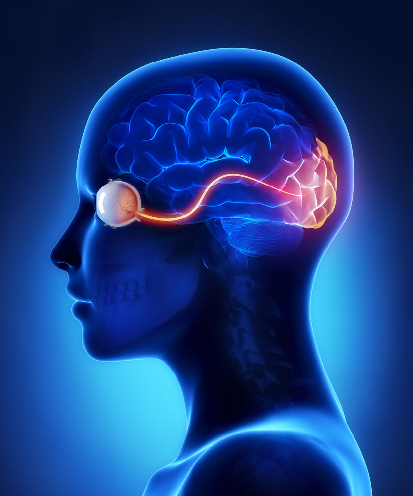
Traumatic optic nerve damage, also referred to as traumatic optic neuropathy (TON), is the result of high-speed collision type injuries that cause closed head injuries and facial trauma and is a significant cause of vision loss in the trauma patient population. Motorcycle and bicycle accidents account for over 60% of cases of TON. Approximately 1 to 5% of patients with closed head injuries with loss of consciousness results in TON. The diagnosis of TON is often delayed because of the impaired mental status in a critically injured ‘polytrauma’ patient that prevents a meaningful eye exam. TON is classified as either ‘Direct TON’ or ‘Indirect TON’.
‘Direct TON’ represents a group of distinctive injuries to the optic nerve diagnosed via MRI or CT scanning or by direct clinical examination by an ophthalmologist. The most devastating injuries that produce immediate and permanent vision loss include optic nerve transection and optic nerve avulsion. Optic nerve avulsion is the situation where the nerve is pulled off the orbit disconnecting the eye from the brain while optic nerve transection is the case whereby the nerve is cut by a bony fragment from facial fractures or skull fractures. Treatable causes of ‘Direct TON’ include an orbital hemorrhage and orbital emphysema. Both may lead to decrease vision from the same mechanism whereby either blood or air builds up behind the eye in the orbit, which is the socket where the eye sits. As blood and air builds up behind the eye, if left untreated, will increase pressures within the orbit that will lead to a decrease in blood flow to the optic nerve and eventual blindness. Orbital hemorrhage is the case where bleeding occurs in the closed space of the orbit and orbital emphysema occurs when there is a fracture of a bone of the orbit and the adjacent sinuses that causes an air pocket. Clinically patients with either orbital hemorrhage or orbital emphysema present with visual impairment, impaired eye movement and bulging of the affected eye produced by the pressure build up behind the eye from either blood or air.
‘Indirect TON’ is believed to be the result of shearing of the nerve fibers of the optic nerve just before it enters the optic canal that occurs in rapid deceleration type injuries. It may also be the result of blunt trauma to the superior part of the orbit or to the lateral forehead. It is believed that the common mechanism of injury to the optic nerve in ‘Indirect TON’ is caused by a compartment syndrome in the optic canal that develops optic nerve swelling in response to nerve damage that will increase the pressures in tight space of the optic canal preventing blood flow to the contained optic nerve. The treatment for ‘Indirect TON’ is controversial at this time and includes the following: observation, high dose steroids, or surgical decompression of the optic canal. Steroids and surgical decompression of the optic canal are utilized to decrease the pressures in the optic canal to improve arterial blood supply to the nerve with a hope that improved blood flow will improve function and prevent further nerve loss. There currently is no consensus in management of ‘Indirect TON’ so risk versus benefits will need to be weighed by the treatment team and generally surgery and steroids are reserved for patients with little to no light perception on exam.
Traumatic orbital injuries are a challenge for the trauma medical team because in the catastrophically injured patient a meaningful physical exam is at best difficult and sometimes impossible because of the impaired mental status. CT scan is the usual choice in evaluating patients with orbital trauma or to those at risk for orbital injuries. Thin-section CT scans can be done very quickly which allows for the diagnosis of orbital fractures, herniation of orbital contents, anterior eye chamber injuries, position of the lens, blood in the eye (hyphema), the presence of foreign bodies, and the appearance of the optic canal. In trauma patients with a rapid decrease in visual acuity, a high-resolution CT scan is necessary to rule out treatable causes of ‘Direct TON’ and view the optic canal. MRI may assist in visualizing an injured optic nerve within the optic canal when determining if high dose steroids or surgical decompression is necessary in the treatment of ‘Indirect TON’.
A trauma ophthalmologist working closely with an Academic Physician Life Care Planner is required in every case of TON since these injuries rarely occur in isolation and there are often associated injuries within the globe that will require future medical care. By using a trauma ophthalmologist to assist with providing the foundation for the life care plan all necessary and appropriate future eye care will be included in the plan understanding that various subspecialties of ophthalmology may be required including glaucoma, retina, and neuro-ophthalmology in the same patient. Neuropsychological assessment must be provided as well considering TON is direct evidence of a likely concomitant traumatic brain injury.
http://content.lib.utah.edu/utils/getfile/collection/EHSL-NOVEL/id/2086/filename/2057.pdf
http://www.nature.com/eye/journal/v18/n11/full/6701571a.html

